Posts
RNA isolation from cells | RNA quantification
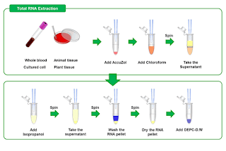
RNA isolation from cells Protocol: Aspirate off media, wash once with ice cold PBS (1-2) mL. Aspirate off PBS, add 1 mL Trizol . Scrape the plate with a cell scraper, collect the cell lysate to 1.5 mL E-tube. Incubate RT for 5mins. Centrifugation 4°C, 13000 rpm for 10 mins. Aliquot supernatant to new E-tube. Add 200 µL chloroform (200 µL + 800 µL=1 mL), vortexing. Incubate RT for 10-15 mins (vortex every 2 mins) Centrifugation 4°C, 13000 rpm for 15 mins. Separate aqueous phase and transfer to new E-tube. Add isopropanol (equal volume of aqueous phase) Inverting, incubate RT for 5-10 mins. Centrifugation 4°C, 13000 rpm for 10 mins. Remove supernatant and add 700 µL 70%EtOH to wash the pellet. Centrifugation 4°C, 13000 rpm for 5 mins. Remove supernatant and add 1 mL 70%EtOH to wash the pellet. Centrifugation 4°C, 7000 rpm for 10 mins. Remove supernatant, spin down. Use yellow tip remove supernatant completely. Air dry rapping with poly for 5-10 mins. Add DE...
Western blot | troubleshooting | samples turn yellow and precipitate

Why western Blot samples turn yellow and precipitate? Sample appears to be acidic. Bromophenol blue in the loading buffer turns to yellow at acidic pH (below 3). Try to adjust the pH by adding NaOH [ 1 ]. Reason for that is proteins may be getting degraded due to heat exposure. One researcher had the same problem and so instead of keeping samples at 95 ° c he started warming at 60-65 ° C. So anyone can try it out and as [ 1 ] said check the pH as well of the loading dye [ 2] . Heating a sample also changes pH. An altered pH is not only responsible to turn the sample color yellow, but may also precipitate the proteins. Keeping SDS containing samples in ice is another common mistake to see this precipitation [ 3 ]. Another researcher said that he has got to do more with the total protein concentration in the lysate. He faced the same problem of precipitation upon denaturation at 100 ° c . He added 1/10th volume of 10% SDS and then boiled. P...
Western blot | Re-use a primary antibody

Why reuse primary antibody? Western blotting is one of the most common method for detecting and semi-quantifying specific proteins. The cost of running a Western is predominantly driven by detection antibodies, which are used to probe membrane-bound target protein(s). Although some antibodies can be produced cheaply in-house, most antibodies must be commercially ordered. Reusing antibodies for subsequent Westerns can save your lab a considerable amount of money. So money is nothing but everything. In conclusion economic research practises is budget friendly and reuse of primary antibody is one of them. When reuse is problematic? The most standard phospho-antibody protocols involve blocking with 5% milk in PBS or TBS (0.1% Tween 20), followed by a couple washing steps with PBS or TBS to remove the excess milk. If you dilute your antibody in a milk solution, storage and re-use will be extremely problematic, because the milk will "go bad" and fall...
Western blot | Bradford assay ⎸Protein quantification

Lab. Bioclock & Aging/Dewan Md. Sumsuzzman Bradford assay ⎸Protein quantification A. Preparation of BSA standard curve Protocol: Preparation of 10 mg/ml BSA solution (10 ml DW + 100 mg BSA) [ Stock solution ] Prepare 1 mg/ml BSA solution (9 ml DW + 1 ml of 10 mg/ml BSA) [ Working solution ] Levelling total 8 cuvettes as Blank, 1, 2, 4, 6, 8, 10, 12 µL respectively. For blank, add 1 mL lysis buffer. And, add 1 mL working solution to 1, 2, 4, 6, 8, 10, 12 µL levelled cuvettes respectively. Discard 1, 2, 4, 6, 8, 10, 12 µL working solution from 1, 2, 4, 6, 8, 10, 12 µL levelled cuvettes gradually. Start quantification and get a BSA curve. B. Protein quantification Protocol: Part-1 (Prepare working solution): Prepare lysis buffer (Western blot protocol) Mix the tissue with lysis buffer in a conical tube (10 mg tissue/1 mL lysis buffer) and incubate for one hour. Prepare bradford working solution by mixing bradford stock solution and autoclaved DW with a r...
Western blot | ECL development
Lab. Bioclock & Aging/Dewan Md. Sumsuzzman ECL development Protocol Remove ECL Western Blotting Reagent Kit (Pierce® ECL plus western blotting substrate) from the refrigerator. Prepare the substrate working solution by mixing Substrate A and Substrate B in a 40:1 ratio (example: 12 mL substrate A + 300 µL substrate B for 8 × 12 cm2 membrane). Use 0.125mL working solution per cm2 of membrane. Note: The working solution is stable for up to 1 hour at RT. Place the membrane on the other suitable clean surface, remove the water, add Pierce® ECL plus western blotting substrate (working solution) and spread evenly over the membrane surface. Incubate blot with working solution for 5 minutes at RT. Remove blot from working solution and place it in a plastic sheet protector or clear plastic wrap. Use an absorbent tissue to remove excess liquid and carefully press out any bubbles from between the blot and the membrane protector. Place the protected membrane in a fil...