Posts

Egg shell | Industrial applications of egg shell

Image
EGGSHELL CHARACTERISTICS: Eggshell waste includes the shell and a membrane that separates the shell from the albumen (the white). These materials consist of as follows: • 94% to 97% calcium carbonate; • 3% to 6% protein; • 1% various minerals (including magnesium, potassium, and traces of iron, sulfur and phosphorus). Eggshell APPLICATIONS AND POTENTIAL MARKETS FOR THE DIFFERENT EGGSHELL COMPONENTS: Emerging sectors for eggshell application Source: https://doi.org/10.1016/j.resconrec.2016.09.027
Stem cells promote peripheral nerve regeneration

This is an embedded Microsoft Office presentation, powered by Office Online .

Patterning factors | determine regional identity and differentiation potential

Ptf1a | Direct reprogramming of fibroblasts into neural stem cells

RNA isolation from cells | RNA quantification

Image
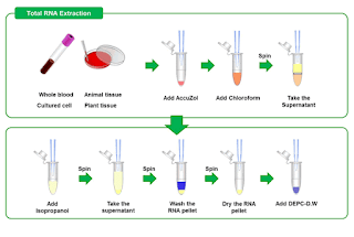
RNA isolation from cells Protocol: Aspirate off media, wash once with ice cold PBS (1-2) mL. Aspirate off PBS, add 1 mL Trizol . Scrape the plate with a cell scraper, collect the cell lysate to 1.5 mL E-tube. Incubate RT for 5mins. Centrifugation 4°C, 13000 rpm for 10 mins. Aliquot supernatant to new E-tube. Add 200 µL chloroform (200 µL + 800 µL=1 mL), vortexing. Incubate RT for 10-15 mins (vortex every 2 mins) Centrifugation 4°C, 13000 rpm for 15 mins. Separate aqueous phase and transfer to new E-tube. Add isopropanol (equal volume of aqueous phase) Inverting, incubate RT for 5-10 mins. Centrifugation 4°C, 13000 rpm for 10 mins. Remove supernatant and add 700 µL 70%EtOH to wash the pellet. Centrifugation 4°C, 13000 rpm for 5 mins. Remove supernatant and add 1 mL 70%EtOH to wash the pellet. Centrifugation 4°C, 7000 rpm for 10 mins. Remove supernatant, spin down. Use yellow tip remove supernatant completely. Air dry rapping with poly for 5-10 mins. Add DE...

Western blot | troubleshooting | samples turn yellow and precipitate

Image
Why western Blot samples turn yellow and precipitate? Sample appears to be acidic. Bromophenol blue in the loading buffer turns to yellow at acidic pH (below 3). Try to adjust the pH by adding NaOH [ 1 ].  Reason for that is proteins may be getting degraded due to heat exposure. One researcher had the same problem and so instead of keeping samples at 95 ° c  he started warming at 60-65 °  C. So anyone can try it out and as [ 1 ] said check the pH as well of the loading dye [ 2] .  Heating a sample also changes pH. An altered pH is not only responsible to turn the sample color yellow, but may also precipitate the proteins. Keeping SDS containing samples in ice is another common mistake to see this precipitation [ 3 ].  Another researcher said that he has got to do more with the total protein concentration in the lysate. He faced the same problem of precipitation upon denaturation at 100 ° c . He added 1/10th volume of 10% SDS and then boiled. P...

Western blot | Re-use a primary antibody

Image
Why reuse primary antibody? Western blotting is one of the most common method for detecting and semi-quantifying specific proteins. The cost of running a Western is predominantly driven by detection antibodies, which are used to probe membrane-bound target protein(s). Although some antibodies can be produced cheaply in-house, most antibodies must be commercially ordered. Reusing antibodies for subsequent Westerns can save your lab a considerable amount of money. So money is nothing but everything. In conclusion economic research practises is  budget friendly and reuse of primary antibody is one of them.  When reuse is problematic? The most standard phospho-antibody protocols involve blocking with 5% milk in PBS or TBS (0.1% Tween 20), followed by a couple washing steps with PBS or TBS to remove the excess milk.  If you dilute your antibody in a milk solution, storage and re-use will be extremely problematic, because the milk will "go bad" and fall...

Western blot | Bradford assay ⎸Protein quantification

Image
Lab. Bioclock & Aging/Dewan Md. Sumsuzzman Bradford assay ⎸Protein quantification A. Preparation of BSA standard curve Protocol: Preparation of 10 mg/ml BSA solution (10 ml DW + 100 mg BSA) [ Stock solution ] Prepare 1 mg/ml BSA solution (9 ml DW + 1 ml of 10 mg/ml BSA) [ Working solution ] Levelling total 8 cuvettes as Blank, 1, 2, 4, 6, 8, 10, 12 µL respectively. For blank, add 1 mL lysis buffer. And, add 1 mL working solution to 1, 2, 4, 6, 8, 10, 12 µL levelled cuvettes respectively. Discard 1, 2, 4, 6, 8, 10, 12 µL working solution from 1, 2, 4, 6, 8, 10, 12 µL levelled cuvettes gradually. Start quantification and get a BSA curve. B. Protein quantification Protocol: Part-1 (Prepare working solution): Prepare lysis buffer (Western blot protocol) Mix the tissue with lysis buffer in a conical tube (10 mg tissue/1 mL lysis buffer) and incubate for one hour. Prepare bradford working solution by mixing bradford stock solution and autoclaved DW with a r...

Western blot | ECL development

Image
Lab. Bioclock & Aging/Dewan Md. Sumsuzzman  ECL development Protocol Remove ECL Western Blotting Reagent Kit (Pierce® ECL plus western blotting substrate) from the refrigerator. Prepare the substrate working solution by mixing Substrate A and Substrate B in a 40:1 ratio (example: 12 mL substrate A + 300 µL substrate B for 8 × 12 cm2 membrane). Use 0.125mL working solution per cm2 of membrane. Note: The working solution is stable for up to 1 hour at RT. Place the membrane on the other suitable clean surface, remove the water, add Pierce® ECL plus western blotting substrate (working solution) and spread evenly over the membrane surface. Incubate blot with working solution for 5 minutes at RT. Remove blot from working solution and place it in a plastic sheet protector or clear plastic wrap. Use an absorbent tissue to remove excess liquid and carefully press out any bubbles from between the blot and the membrane protector. Place the protected membrane in a fil...

Western blot | Antibody incubation & Washing

Image
Lab. Bioclock & Aging/Dewan Md. Sumsuzzman Antibody incubation & Washing ※ When you first take the Antibody, it tells you how much concentration should be diluted. Use it at that concentration. If the protein band comes out, continue to use it. If the band does not come out, increase the concentration. Primary & secondary antibody incubation ※ If there are several proteins you want to see, the membrane may be cut to size. Prior to Primary Ab treatment, remove the water, wrap it in a wrap, and check for protein markers (total volume 2-3 ml). A. Dilution of primary Ab: Dilute primary Ab in TBST (0.5% Skim milk / TBST) and incubate shaking incubation for 1 hour at room temperature. Or overnight at 4 ° C. → 0.5% skim milk + TBST + primary Ab → Used primary Ab can be re-used, can be kept at 4 ℃ Ex 1) TBST 10 ml + Skim milk 0.05 (g) + 10 μl of primary AB when the dilution concentration is 1: 1000 and the total volume is 10 ml Ex 2) TBST 2 ml + ...

Western blot | Protein transfer

Image
Lab. Bioclock & Aging/Dewan Md. Sumsuzzman Transfer membrane Preparation: • 10X & 1X Transfer buffer (4℃) • Sponge / Filter paper / 3MM paper / Membrane • Cassette / Blotting chamber • 10X TBS (pH 7.5) &  1X TBST • 0.2% Ponceau S • Coomassie blue • Blocking buffer (5% skim milk in TBST) • Staining solution & Destaining solution • BM purple AP substrate Transfer protocol: Cut the membranes to the appropriate size and immerse them in transfer buffer. For PVDF membrane, immerse in methanol for 5 minutes before transferring to transfer buffer. Membrane activation:  The process by which proteins in the gel make hydrophobic interaction with the membrane. This process is not necessary when using an NC. Transfer the electrophoresis gel to the transfer buffer, remove the wells, and remove the remaining portion except the sample. → Save membrane. Place the black portion of the Western blot cassette against the bot...

Western blot | Ponceau S Staining

Image
Lab. of Bioclock & Aging/Dewan Md. Sumsuzzman Ponceau S Staining Ponceau S Staining Solution: Table-1: Recipe of 0.1%  (w/v) Ponceau S in 5% (v/v) Acetic acid Ingredients Quantity Ponceau S 1 gm Acetic acid 50 ml ddH2O Make up to 1L Table-2: Recipe of 0.2%  (w/v) Ponceau S in 3% (v/v) Acetic acid Ingredients Quantity Ponceau S 2 gm Acetic acid 30 ml ddH2O Make up to 1L Table-3: Recipe of 0.1N NaOH in 100 ml water Ingredients Quantity NaOH 0.4 gm H2O 100 ml Protocol: Following Western blotting, transfer the membrane to 5ml Ponceau S Stain solution. Place on an orbital shaker for 5 minutes at room temperature. Rinse membrane with DI water to achieve desired staining, approximately 2‐3 washes of 5 minutes each will remove the background staining. For total protein destaining, use a 0.1N NaOH solution. Wash the membrane with 0.1N NaOH solution for 5 minutes. Protein bands will start disappear after 10-30 seconds. Discard wash solution and repeat once. Wash the membrane...

Western blot | Coomassie Blue Staining

Image
Lab. Bioclock & Aging/Dewan Md. Sumsuzzman Coomassie Blue Staining Table-1: Recipe of Staining Solution Ingredients Quantity Coomassie Blue R-250 2.5 gm Methanol 500 ml Acetic acid 100 ml DW 400 ml Note:  Final volume 1L. Add 100 ml of acetic acid to 500 ml of ddH2O. Add 500 ml of methanol and mix. Add 1g of Coomassie R250 dye and mix.  Put a magnetic bar in a brown bottle, and add dye while spinning. Finally, filter it. Store at room temperature in a sealable container. Table-2: Recipe of Destaining Solution Ingredients Quantity Methanol 500 ml Acetic acid 100 ml DW 400 ml Store at room temperature in a sealable container. Protocol After the gel has finished running, remove it from the tank and remove the gel from the glass plates. Wash the gel 2-3 times, 5 minutes each in deionized water to remove SDS present in the gel. Each wash should be large volumes of water. Remove all free water from the gel. Add an adequate amount of staining solution (Coomassie Blue R-250) to ...